Department of Histology and Embryology – Faculty of Medicine, Masaryk University
MUDr. Jana Dumková, Mgr. Marek Stehlík
Introduction
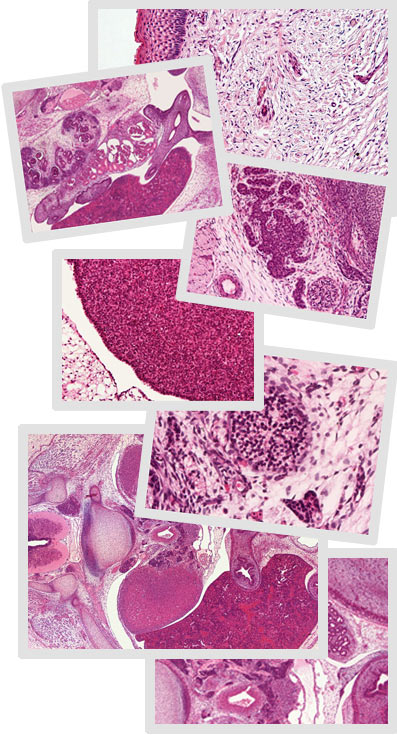
This interactive atlas of human embryology is an image supplementation of the learning texts from the embryology. The atlas recapitulates prenatal human development and observes the changes which are happening during the first days and weeks after the successful fertilization. The original micrographs presented in the atlas are briefly labeled to reach the better lucidity and an understanding of developmental changeovers proceeding in human. The interactive atlas of human embryology is divided into following chapters.
The first chapter is exceptional because it is devoted to the beginnings of human development. It is divided into three parts – the first part is aimed to the organs in which germ cells are developing, and then to the female and male germ cells. At the second part, the processes of fertilization, the cleavage of an oocyte, implantation of the blastocyst and also the differential processes occurring in the blastocyst during the 3rd week of development are described. The third part is aimed to the morphology of accessory embryonic organs, i.e. morphology of placenta and umbilical cord.
The next chapters of this atlas are arranged according to the organ systems – include development of the heart and the large vessels; of the thymus and the spleen; of the respiratory system; of the digestive system; of the urinary system and the genital system, also development of the endocrine system; of the nerve system; of the eye and the ear and naturally also of the integumentary system.
Quite exceptional is also the last chapter, because documents the development of limbs and contains the images dedicated to the chondrogenic ossification of hand bones.
All chapters of this interactive atlas of embryology contain the histological sections especially of human embryos; the micrographs are arranged subsequently from the 6th week of embryonic development to the 9th week of development, when we begin to speak about the human fetus; in some organ systems the images from later fetal period are added (the 4th and the 6th month of development).
For better understanding, in some systems are incorporated the images of histological slides coloured by a special staining methods designed especially for an embryonic tissues as e.g. TRIPARS, trichrome green staining, AZAN or staining by methyl green-pyronin.
For the comparison, at the ends of the chapters are inserted also the micrographs of the animal sections – laboratory mouse or laboratory rat, which correspond to the same period of development as in the human embryo.
All images are arranged craniocaudally to capture all important organs of submitting system and to visualize the topographic relations among individual organs. Some slides are proposed in different magnifications; higher magnifications are used in case some fine morphological details have appeared during the specific period of development and these details are characteristic for development of the organ.
The interactive atlas of human embryology is designed for the students of Master's degree programmes of Faculty of Medicine (students of general medicine and dentistry), students of Bachelor's degree programmes of Faculty of Medicine (especially nurses - midwifes), students of doctoral study programmes and medical doctors of some specializations in graduate programmes.
The interactive atlas of human embryology is specialized for the teaching of students focused on natural science and aim to improve the knowledge from prenatal period of human, improve the visualization and understanding of too complicated processes, which are happening before the human birth. Nevertheless I would be pleased if this atlas could be used not only by professionals, but also by general public.
All pictures are chosen to engage your attention by their uniqueness and they are so attractive that they can meaningfully ‘popularize’ the embryology and to increase an interest in this branch of science.
Department of Histology and Embryology, Faculty of Medicine MU |
Back to the homepage, accessibility |
Centrum interaktivních a multimediálních studijních opor pro inovaci výuky a efektivní učení | CZ.1.07/2.2.00/28.0041